Bacteria Cell Vector Art, Icons, and Graphics for Free Download
The three main shapes of bacteria are coccus, spiral, and bacillus. Cocci are bacteria that are spherical or ovoid in shape. Some cocci remain attached after binary fission, even though separate cells have been formed. For example, diplococci are cocci in pairs, streptococci are chains, and staphylococci are clusters of multiple cocci.
Bacteria Diagram Photograph by Monica Schroeder
What are prokaryotes? Prokaryotes are microscopic organisms belonging to the domains Bacteria and Archaea, which are two out of the three major domains of life. (Eukarya, the third, contains all eukaryotes, including animals, plants, and fungi.) Bacteria and archaea are single-celled, while most eukaryotes are multicellular.
Bacterial Structure Plantlet
bacteria, any of a group of microscopic single-celled organisms that live in enormous numbers in almost every environment on Earth, from deep-sea vents to deep below Earth's surface to the digestive tracts of humans. Bacteria lack a membrane-bound nucleus and other internal structures and are therefore ranked among the unicellular life-forms.
Anatomy of Bacteria stock vector. Illustration of labelled 43965779
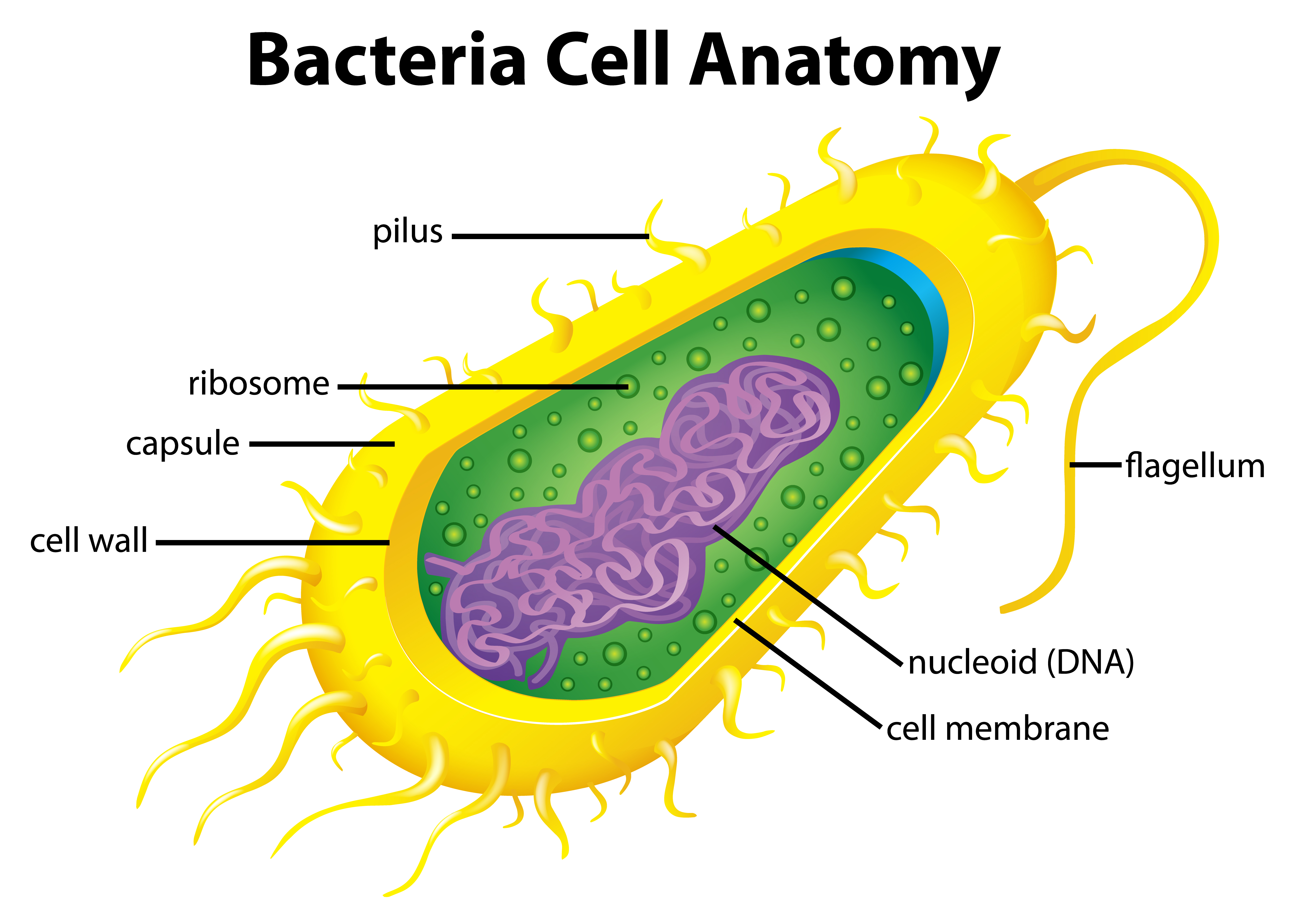
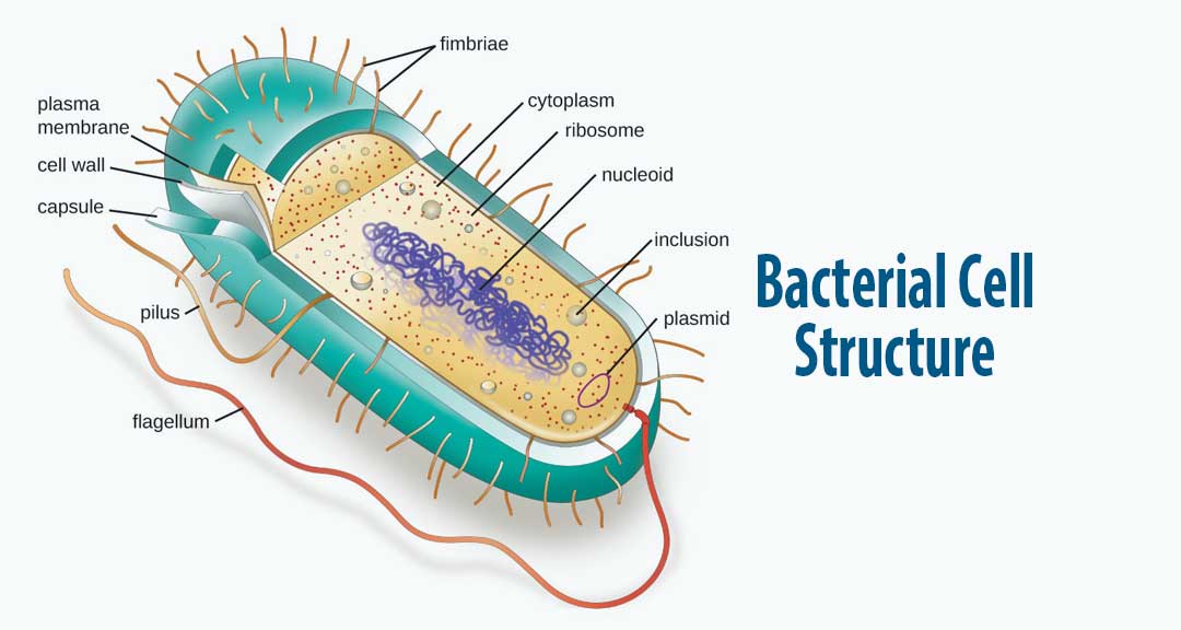
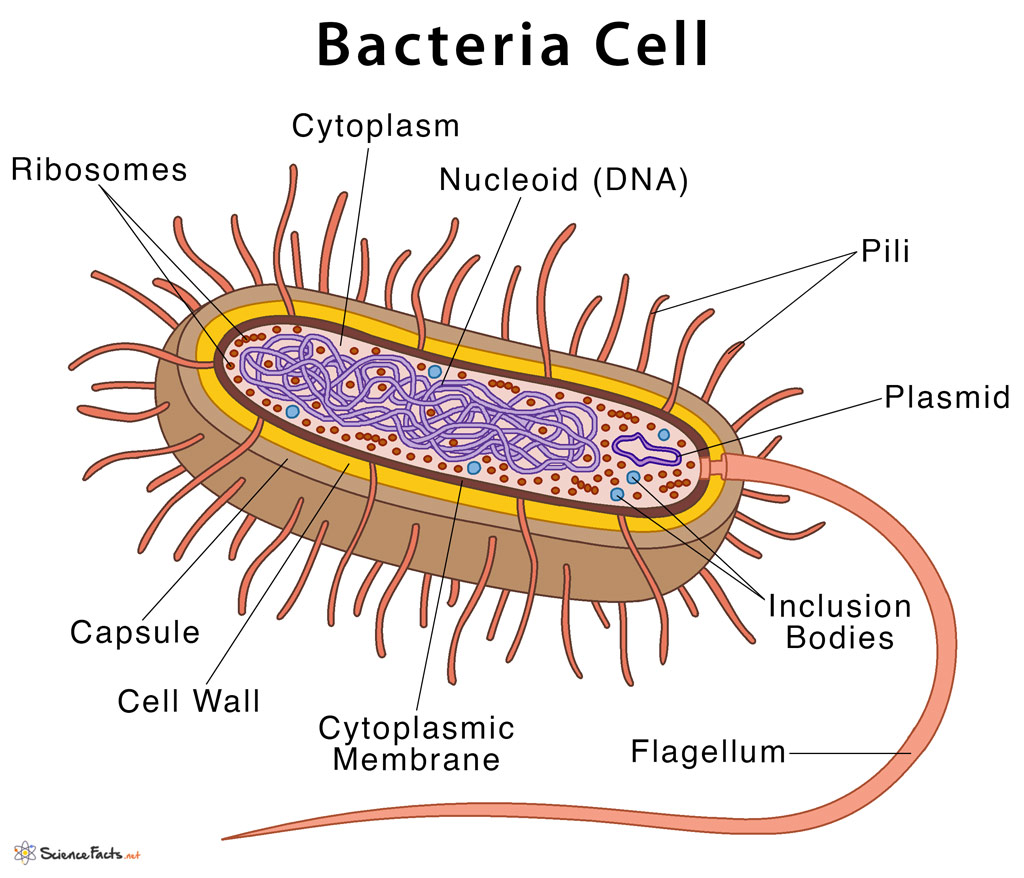
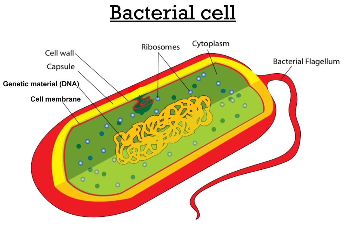
In this article we will discuss about the cell structure of bacteria with the help of diagrams. A bacterial cell (Fig. 2.5) shows a typical prokaryotic structure. The cytoplasm is enclosed by three layers, the outermost slime or capsule, the middle cell wall and inner cell membrane. The major cytoplasmic contents are nucleoid, plasmid, ribosome.
Bacteria Labeled Stock Illustrations 262 Bacteria Labeled Stock Illustrations, Vectors
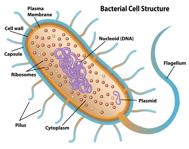
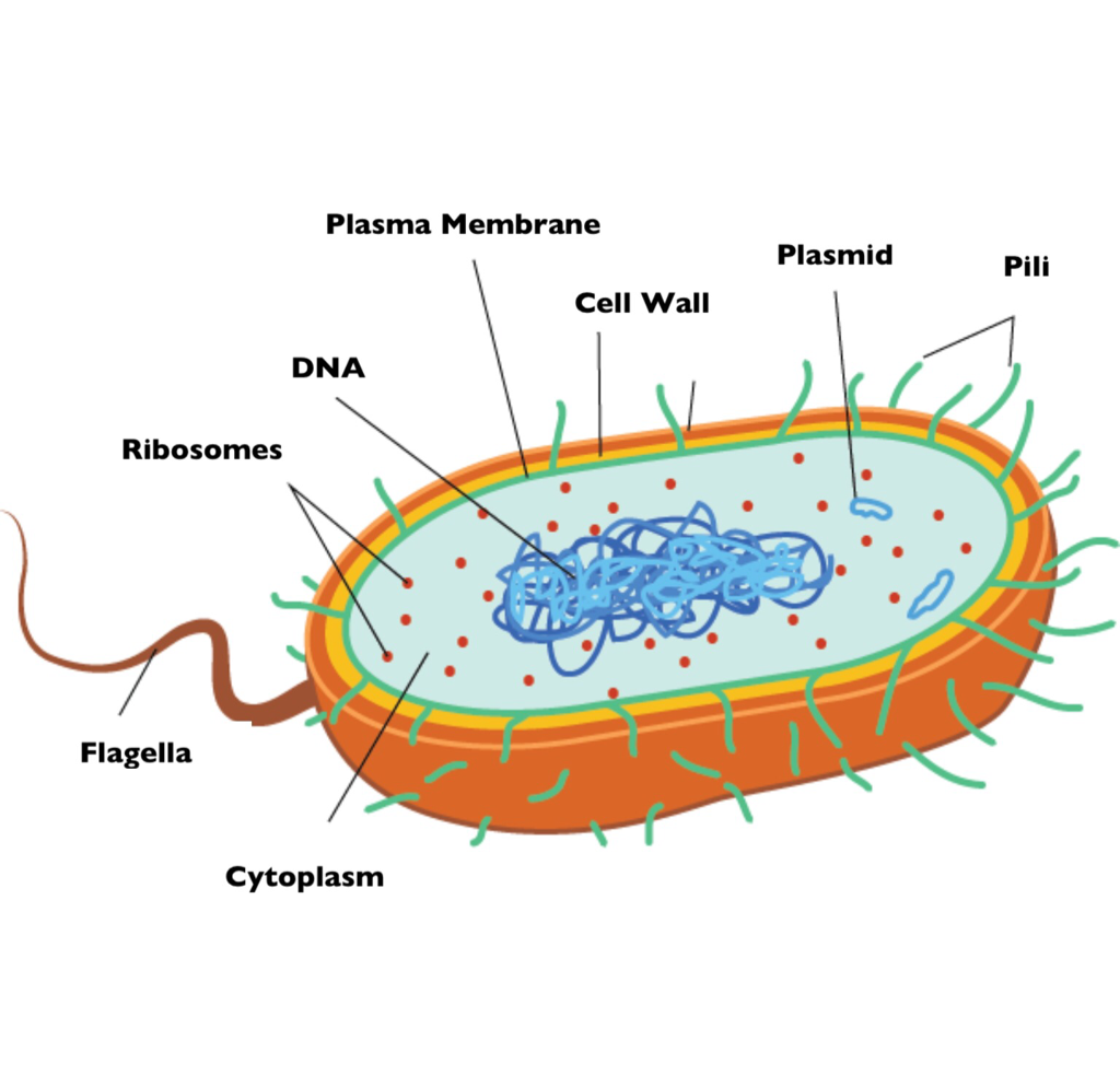
Bacteria Diagram with Labels Bacterial cells have simpler internal structures like Pilus (plural Pili), Cytoplasm, Ribosomes, Capsule, Cell Wall, Plasma membrane, Plasmid, Nucleoid, Flagellum, etc. Labeled Bacteria diagram Eukaryotes have been shown to be more recently evolved than prokaryotic microorganisms.
Bacterial Cell Diagrams 101 Diagrams

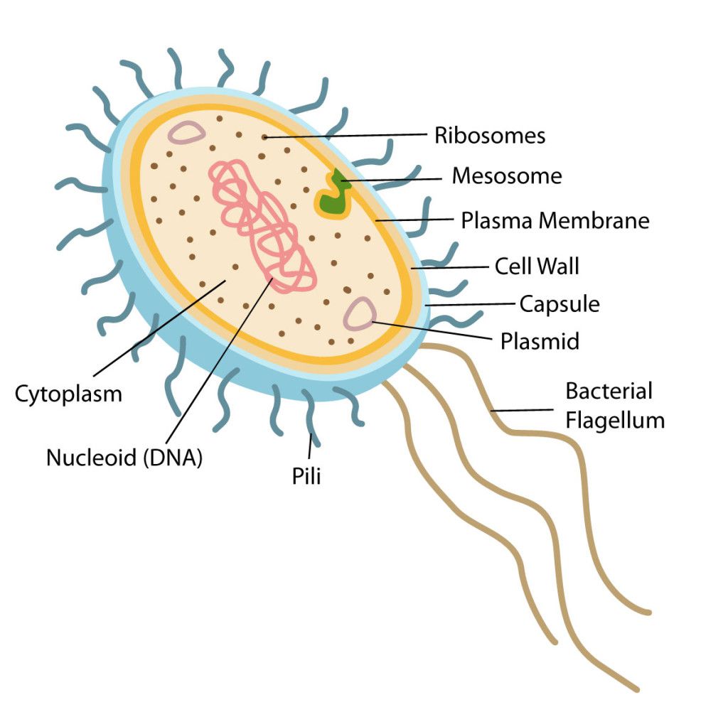
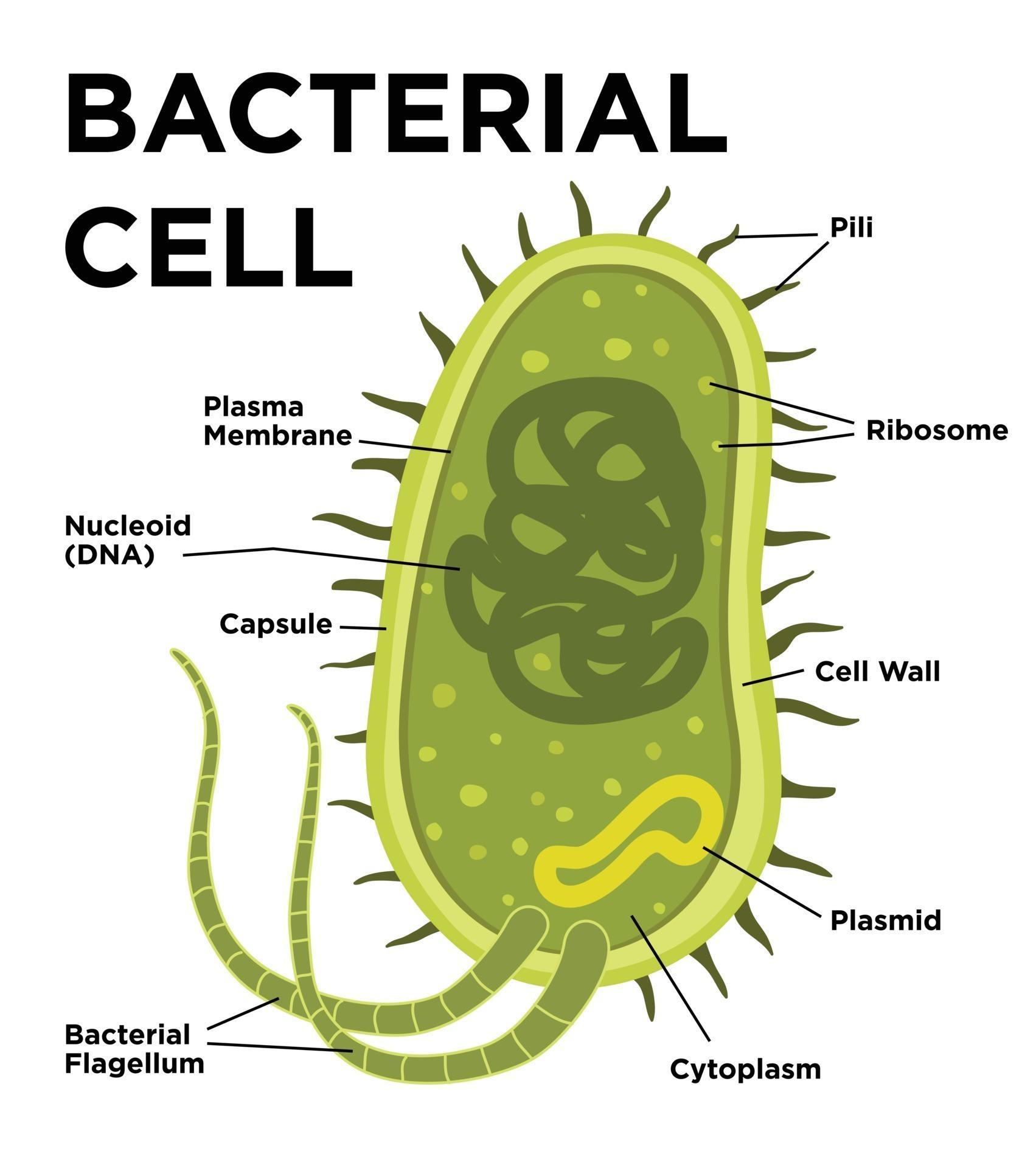
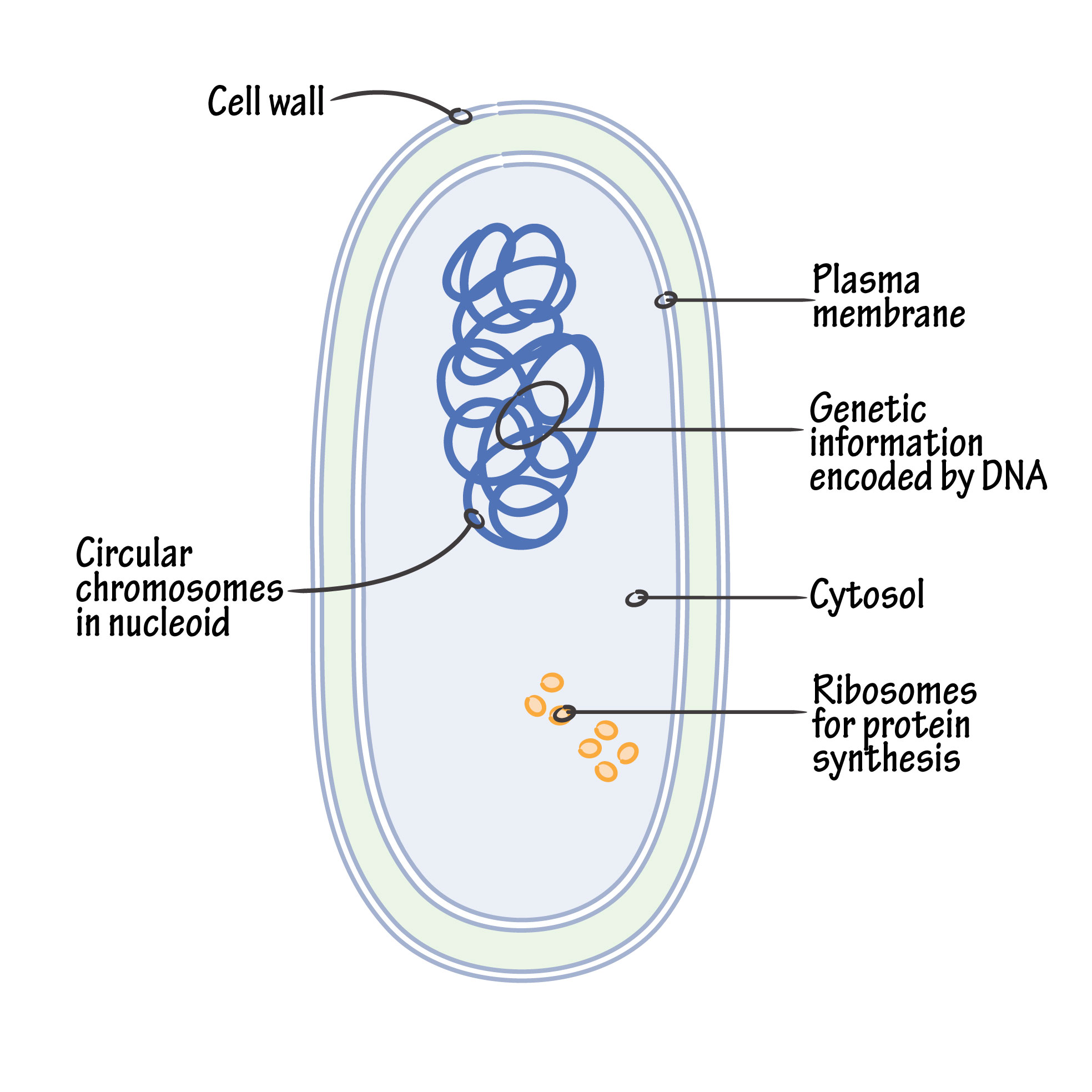
Figure: Labelled diagram of a typical bacterial cell Cell wall. The thick erect elastic membrane that lies beneath the slime layer outside the bacterial cell is called the cell wall. Its thickness is around 10-25 nm and is made up of proteins, lipids, and carbohydrates. Usually, the cell wall does not contain cellulose.
Bacteria Labeled Diagram Stock Vector Art & Illustration, Vector Image 78697031 Alamy
Cell Envelope - The cell envelope is made up of two to three layers: the interior cytoplasmic membrane, the cell wall, and -- in some species of bacteria -- an outer capsule. Cell Wall - Each bacterium is enclosed by a rigid cell wall composed of peptidoglycan, a protein-sugar (polysaccharide) molecule. The wall gives the cell its shape and.
Bacterial Cell Structure and Function
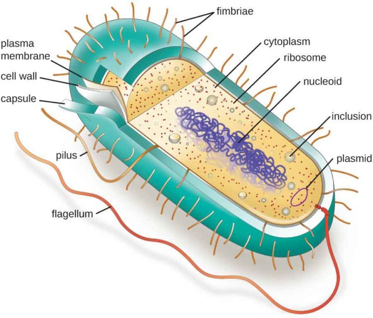
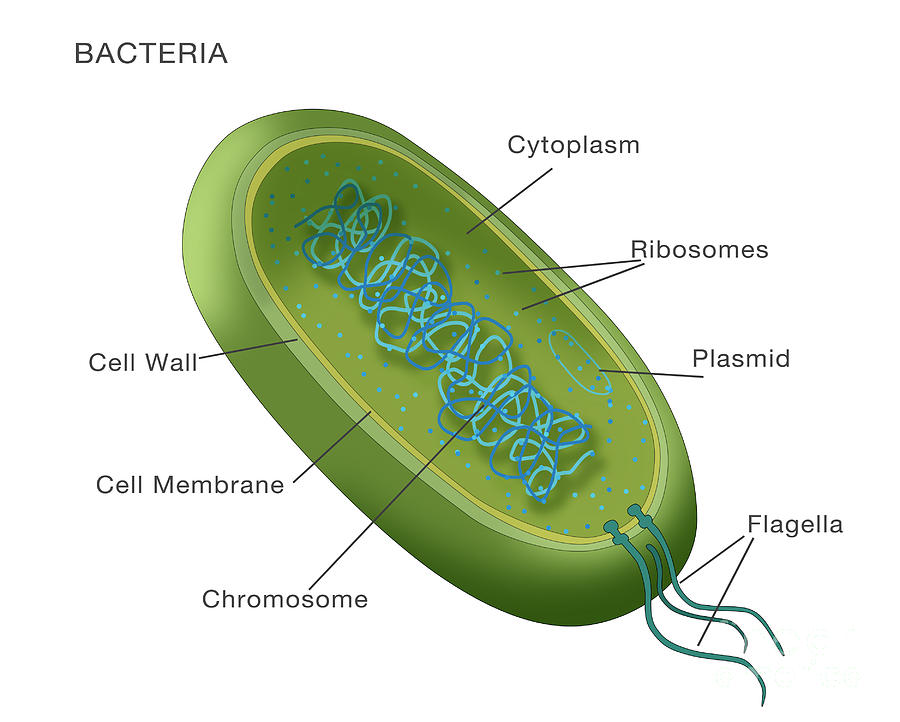
Bacteria Diagram The bacteria diagram given below represents the structure of a typical bacterial cell with its different parts. The cell wall, plasmid, cytoplasm and flagella are clearly marked in the diagram. Bacteria Diagram representing the Structure of Bacteria Ultrastructure of a Bacteria Cell
Bacterial cell anatomy in flat style. Vector modern illustration. Labeling structures on a
coccus (circle or spherical) bacillus (rod-like) coccobacillus (between a sphere and a rod) spiral (corkscrew-like) filamentous (elongated) Cell shape is generally characteristic of a given bacterial species, but can vary depending on growth conditions.
Bacterial Cell Structure and Function
It is a tough and rigid structure of peptidoglycan with accessory specific materials (e.g. LPS, teichoic acid etc.) surrounding the bacterium like a shell and lies external to the cytoplasmic membrane.
Structure of Bacteria Cell and its Organelles Study Wrap

Label a Bacteria Cell Bacteria are tiny single-celled organisms that are all around us, too small to be seen without a microscope. They're living things that come in different shapes like spheres (called cocci), rods (called bacilli), or spirals. Some of them are helpful, like the ones that help us digest food, while others can make us sick.
prokaryotic cell bacteria parts

All bacteria, both pathogenic and saprophytic, are unicellular organisms that reproduce by binary fission. Most bacteria are capable of independent metabolic existence and growth, but species of Chlamydia and Rickettsia are obligately intracellular organisms. Bacterial cells are extremely small and are most conveniently measured in microns (10-6 m). They range in size from large cells such as.
Bacterial Structure Plantlet
1. A bacterial cell remains surrounded by an outer layer or cell envelope, which consists of two components - a rigid cell wall and beneath it a cytoplasmic membrane or plasma membrane. ADVERTISEMENTS: 2.
Characteristics of bacterial cells
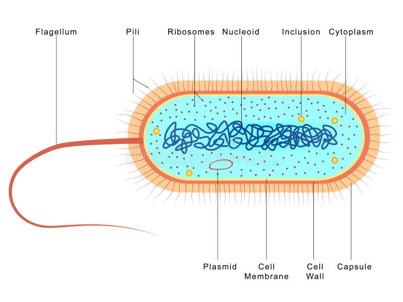
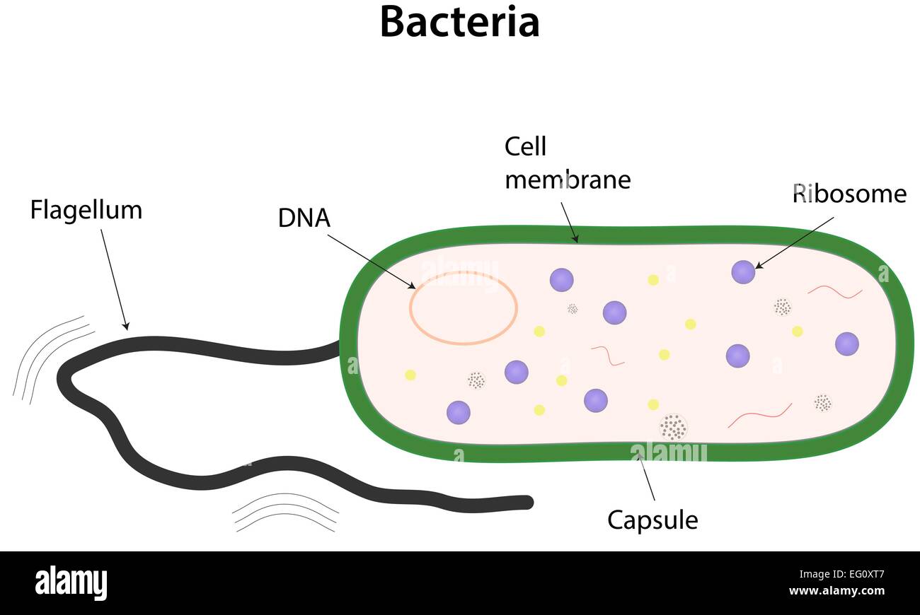
Summary edit. English: A simple diagram of a bacterium, labelled in English. It shows the cytoplasm, nucleoid, cell membrane, cell wall, mitochondria (which bacteria lack), plasmids, flagella, and cell capsule. The SVG code is valid. This diagram was created with an unknown SVG tool.
Bacteria Grade 11 Biology Study Guide
A prokaryote is a simple, single-celled organism that lacks a nucleus and membrane-bound organelles.
Bacteria Ms A Science Online
Ultrasmall Bacteria. Ultrasmall bacteria (150 could fit in a single Escherichia coli) have been discovered in groundwater that was passed through a filter with a pore size of 0.2 micrometers µm). They showed an average length of only 323 nanometers(nm) and an average width of 242 nm. They contain DNA, an average of 42 ribosomes per bacterium, and possessed pili .