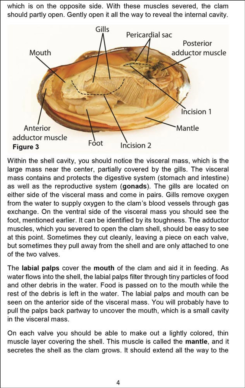
Anatomy Of A Clam
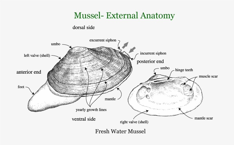
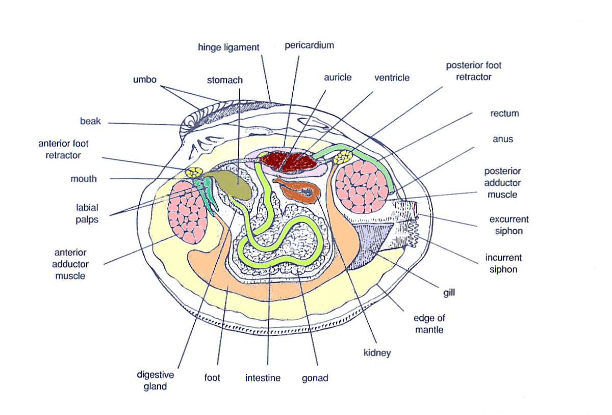
The clam's foot is used to dig down into the sand, and a pair of long siphon s that extrude from the clam's mantle out the side of the shell reach up to the water above (only the exit points for the siphons are shown). Note: this image is colored to differentiate internal organs and are not the actual colors of the clam. Clams are filter feeders.
Clam Dissection BIOLOGY JUNCTION
The style of citing shown here is from the MLA Style Citations (Modern Language Association). When citing a WEBSITE the general format is as follows. Author Last Name, First Name (s). "Title: Subtitle of Part of Web Page, if appropriate." Title: Subtitle: Section of Page if appropriate. Sponsoring/Publishing Agency, If Given.
Clam Dissection Labeled ubicaciondepersonas.cdmx.gob.mx
See a clam diagram, study the clam digestive system read about the excretory system of these animals from the phylum Mollusca. Updated: 11/21/2023 Table of Contents What Is a Clam? Parts of.
Figure 33.21 Anatomy of a clam
Start studying Clam Anatomy. Learn vocabulary, terms, and more with flashcards, games, and other study tools.
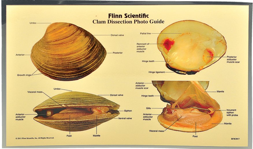
Clam Dissection Guide
Anatomy of a Clam Grade Level: 5-12 Subject Area: Biology, Anatomy Time: Preparation: 10 minutes Activity: 30-45 minutes Clean-up: 10 minutes Student Performance Standards (Sunshine State Standards): 10.03 List examples of aquatic crops and animals (LA.910.1.6.1, 2, 3, 4, 5; LA.910.2.2.2; SC.912.L.17.9).
bioweb images Marine biology, Anatomy, Fish anatomy

Clam dissection is a fantastic first step into the world of anatomy and dissection, particularly for young learners. Clams are easy to find either in the sand or in the supermarket and the project can be looped in with dinner. Learn about clams, dissection, and anatomy in this fun afternoon project.
Clam Diagram Labeled
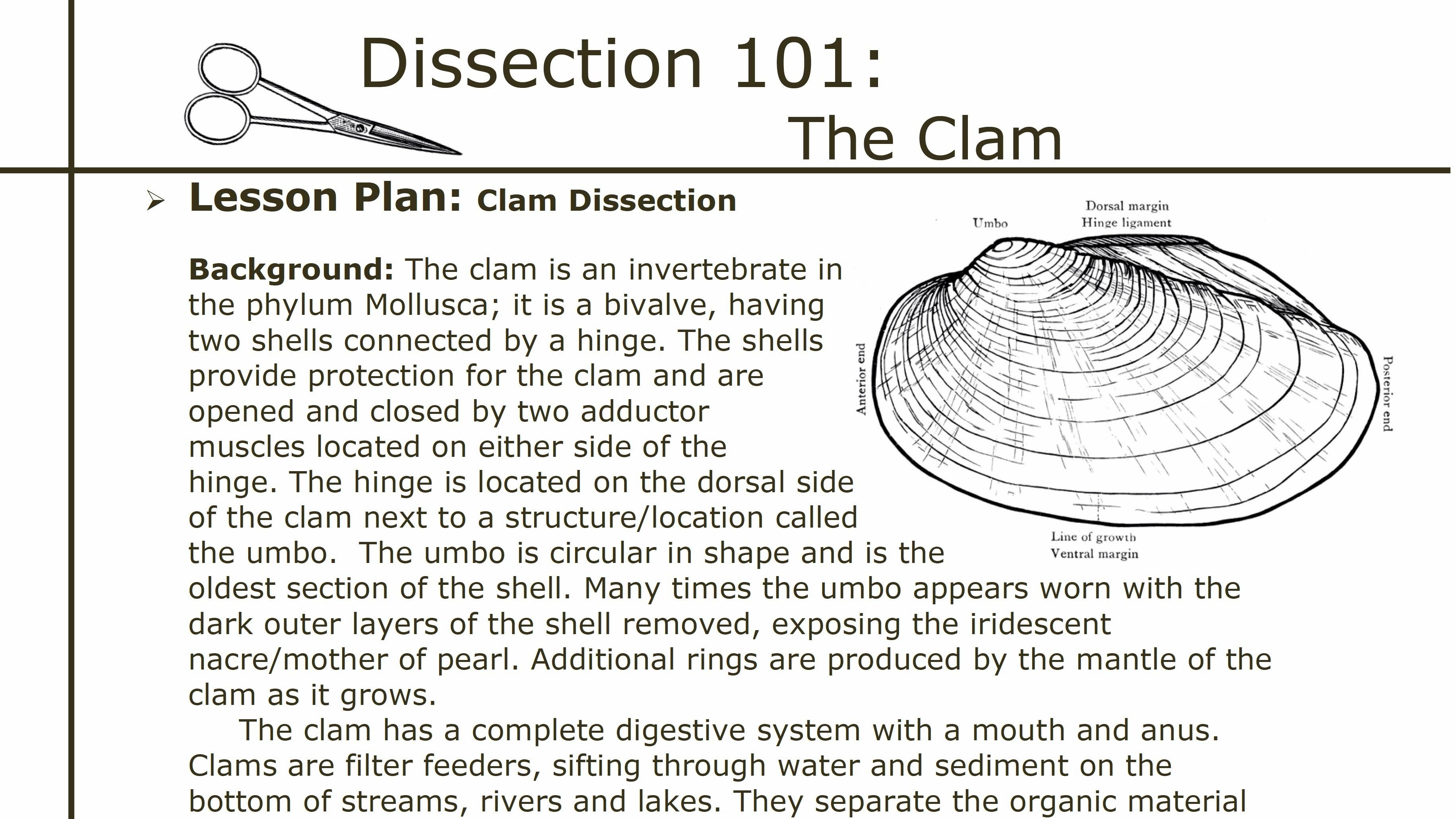
Clam. Clam is a common name for several kinds of bivalve molluscs. The word is often applied only to those that are edible and live as infauna, spending most of their lives halfway buried in the sand of the seafloor or riverbeds. Clams have two shells of equal size connected by two adductor muscles and have a powerful burrowing foot. [1]
Dissection 101 Clam Dissection Lesson Plan PBS LearningMedia
The heart of a clam can be seen in the photograph below. Bivalves have three pairs of ganglia but do not have a brain. Most mollusks have separate sexes but most snails (gastropods) are hermaphrodites.. See the diagram below for the location of the adductor muscles. Figure 4. Adductor muscles of a clam.
Clam Dissection Diagram Quizlet
Snapshot: Bivalvia. Phylum Mollusca, Class Bivalvia. Common names of representatives: clams, scallops, oysters, mussels. Habitat(s): marine (salt water), freshwater (lakes, rivers, and streams). Feeding type(s): mostly suspension feeders; some deposit feeders and carnivores Geological range: Cambrian to today. Clade defining feature(s): two hinged shells surrounding a body with large stomach.
anatomy of a clam Marine biology, Clams, Anatomy

This video details the external and internal anatomy of a clam. Additional video, lesson plans, quizzes, additional dissections, and more are available in the in the Support Materials section above and in the Dissection 101 Collection.
Clams Anatomy Barnegat Bay Shellfish
C. Would you rather study a diagram to learn about a clam or investigate a real clam? Explain. Posterior Adductor Muscle Scar DORSAL Hinge Ligament Drawings courtesy of BIODIDAC. SIDE VENTRAL SIDE Anterior Adductor Muscle Scar Pallial Line (attachment of mantle) Outside of the clam: 1). The soft body is protected by how many shells (valves)?
Clam Anatomy Diagram Quizlet

The diagram below provides an overview not only of that first year of growth, but also of the full softshell clam lifecycle from starting as an egg through adulthood and reproduction. Lifecycle of the soft shell clam (reproduced from a 1983 Maine Department of Resources report by C.T. Newell)
Clam Anatomy Diagram

Clam Dissection Lab: Explained. The phylum Mollusca includes snails, clams, chitons, slugs, limpets, octopi, and squid. As mollusks develop from a fertilized egg to an adult, most pass through a larval stage called the trocophore. The trocophore is a ciliated, free-swimming stage. Mollusks also have a radula or file-like organ for feeding, a.
Clam Anatomy Diagram anatomy diagram source

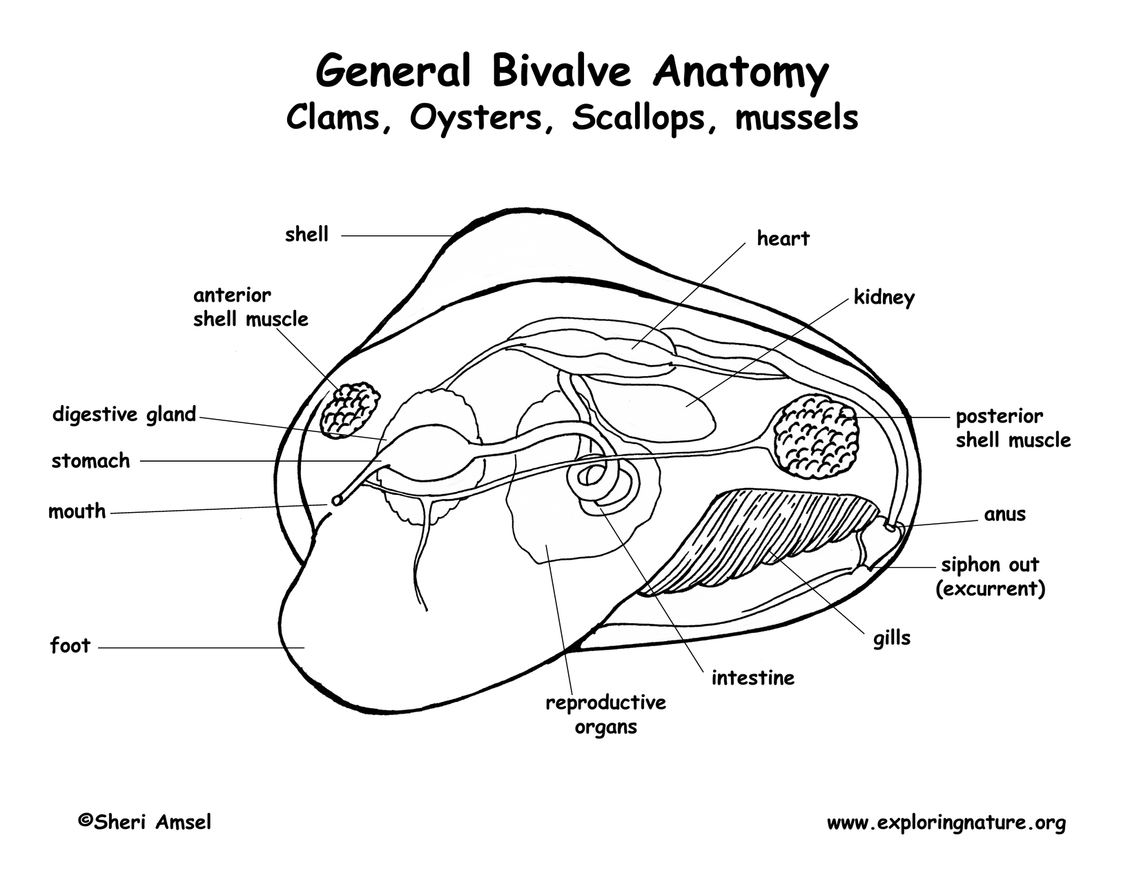
Internal Clam Shell Anatomy 1. Mantle •Covers visceral or body mass •Holds in fluid •Secrets new shell 2. Ant. adductor muscle 3. Post. adductor muscle •Hold valves shut 4. Pericardium cavity •Region covered with thin, dark membrane •Contains 2-chambered heart and kidney in a fluid-filled sac 5. Mantle edge
Clams Characteristics, properties, reproduction and more

Location Cardinal Teeth of Clam a tooth of the hinge of a bivalve mollusk's shell situated just under the umbo and often relatively large Gonad of Clam A sex gland or reproductive gland Mantle of Clam
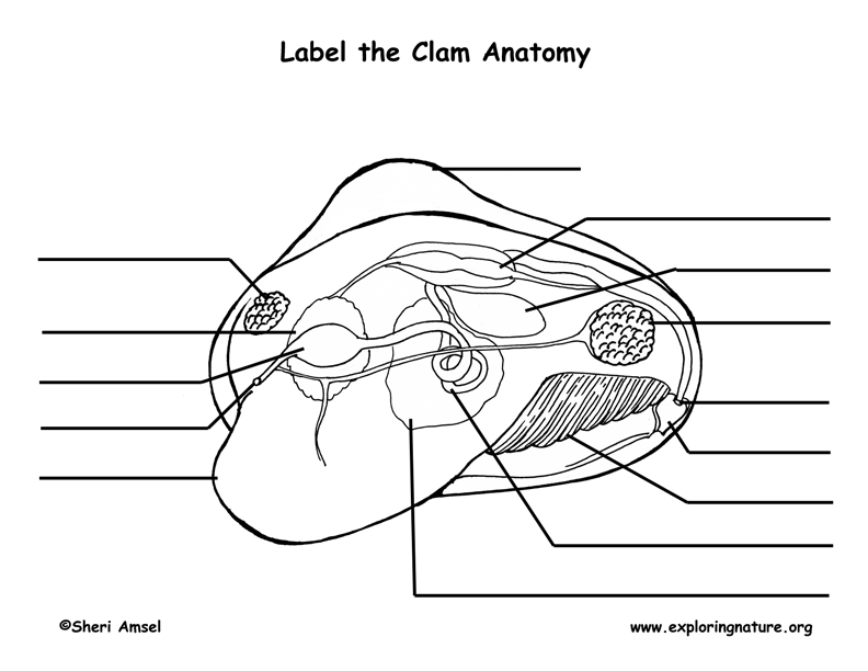
Clam Anatomy Labeling Page
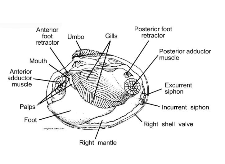
Clams are marine mollusks with two valves or shells. Like all mollusks, a clam has a mantle which surrounds its soft body. It also has a muscular foot which enables the clam to burrow itself in mud or sand. The soft tissue above the foot is called the visceral mass and contains the clam's body organs. Taxonomy Kingdom - Animalia Phylum - Mollusca